2013年5月23日星期四

question 159-169

Vit B12 deficiency in  diabetic patients
Clinical findings – distal axonopathy (numbness) + central myelopathy(loss of position sense)
Lab findings – anemia (megaloblastic) + HB a1c < 7 (good glycemic control)
Loss of position sense is an early sign of vitamin B12 deficiency. 
What is the most likely cause? Every option has an association with Vitamin B 12 deficiency (they are not direct causes of the deficiency) but only chronic metformin use is a direct cause. Metformin reduces vitamin B12 absorption by inhibiting calcium dependent ileal membrane transport of this vitamin. Vitamin B12 and calcium supplementation is the only effective way to alleviate the symptoms of central myelopathy.
Diabetic patients who develop loss of joint position sense should be treated with Vitamin B 12. Thyroid deficiency does not directly cause central myelopathy. Chronic metformin use is an independent cause of Vitamin B 12 deficiency that should be treated with Vitamin B 12 & calcium supplements. 

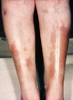
Complex regional pain syndrome (CRPS)

  • severe burning pain
  • pathological changes in bone and skin
  • excessive sweating
  • tissue swelling
  • extreme sensitivity to touch
If complex regional pain syndrome isn't diagnosed and treated early, the disease may progress to more disabling signs and symptoms. These may include:
  • Tissue wasting (atrophy). If you avoid moving an arm or a leg because of pain or if you have trouble moving a limb because of stiffness, your skin, bones and muscles may begin to deteriorate.
  • Muscle tightening (contracture). You may also experience tightening of your muscles. This may lead to a condition in which your hand and fingers or your foot and toes contract into a fixed position.
treatment
Medications Doctors use various medications to treat the symptoms of complex regional pain syndrome.

  • Pain relievers. Over-the-counter (OTC) pain relievers, such as aspirin, ibuprofen (Advil, Motrin, others) and naproxen (Aleve), may ease pain and inflammation. 
  • Antidepressants and anticonvulsants. Sometimes antidepressants, such as amitriptyline, and anticonvulsants, such as gabapentin 
  • Corticosteroids. Steroid medications, such as prednisone, may reduce inflammation and improve mobility in the affected limb.
  • Bone-loss medications. Your doctor may suggest medications to prevent or stall bone loss, such as alendronate (Fosamax) and calcitonin (Miacalcin).
  • Sympathetic nerve-blocking medication. Injection of an anesthetic to block pain fibers in your affected nerves may relieve pain in some people.

Therapies
  • Applying heat and cold. Applying cold may relieve swelling and sweating. If the affected area is cool, applying heat may offer relief.
  • Topical analgesics. Various creams are available that may reduce hypersensitivity, such as lidocaine or a combination of ketamine, clonidine and amitriptyline.
  • Physical therapy. Gentle, guided exercising of the affected limbs may help decrease pain and improve range of motion and strength. The earlier the disease is diagnosed, the more effective exercises may be.
  • Transcutaneous electrical nerve stimulation (TENS). Chronic pain is sometimes eased by applying electrical impulses to nerve endings.
  • Biofeedback. In some cases, learning biofeedback techniques may help. In biofeedback, you learn to become more aware of your body so that you can relax your body and relieve pain.
  • Spinal cord stimulation. Your doctor inserts tiny electrodes along your spinal cord. A small electrical current delivered to the spinal cord results in pain relief.
Dressler’s syndrome is typically treated with NSAIDs such as aspirin or with corticosteroids.[9] However corticosteroids are reserved for rare cases and are seldom required.

Osteonecrosis of the Hip


Diagnosis and treatment
It is estimated that doctors see about 10,000-20,000 new cases of osteonecrosis (ON) each year. No one knows exactly what causes it. See your doctor if you start feeling a dull ache or throbbing pain to the side of your hip in the groin or buttock and you have osteonecrosis risk factors including:
  • Age 20-50 years.
  • Hip dislocation or fracture.
  • Alcoholism.
  • Corticosteroid use.
  • Glandular problems and diseases including rheumatoid arthritis, sickle cell disease, myeloproliferative disorders, Gaucher's disease, chronic pancreatis, Crohn's disease, Caisson's disease or systemic lupus erythematosus.
Your doctor may flex and rotate your hips to check for pain. Your hips may be X-rayed and possibly scanned by MRI (magnetic resonance imaging) to see if bone marrow is dying or dead, and how much the head of your femur may have collapsed.
  • If you have ON and the head of your femur is not yet collapsed, certain medical procedures (i.e.: decompression and bone grafting) may help your body build new blood vessels and bone cells to replace the dead ones.
  • If ON has already collapsed your hip, total hip replacement surgery (arthroplasty) may eliminate your pain and give you better hip mobility. A ball and socket replaces your hip joint. Your thighbone is fitted with the ball piece, which takes the place of the head of your femur. Your hip socket is fitted with the socket piece (cup).

2013年5月21日星期二

observership 2

Atrial fibrillation CHADS(2) score for stroke risk
C congestive heart failure 1
H  hypertension above 140/90  1
A >75                             1
D DM                             1
S previous stroke/TIA/thromboembolism  2

score                          Risk                           anticoagulation therapy
0                                  low                               none or aspirin daily
1                                 moderate                       aspirin or wafarin, aspirin daily or INR 2-3
2 or greater                 moderate or high            wafarin, raise INR 2-3 unless contraindication

OA joint change


asthma exacerbation
detecting the onset of an exacerbation
decrease in peak flow of greater 20% from normal  signals the deterioration
a fall to less than 50% of baseline signals severe attack
treatment:
  • duoneb (inhaled albuterol/ipratropium)
  • systemic glucocorticoids, for patients w/impending respiratory failure, give methylprednisolone 60-125 mg IV, for majority of less severe, give prednisolone 40-60 mg orally
  • continue advair (Fluticasone and salmeterol, Beta;2 Agonist Beta;2-Adrenergic Agonist, Long-Acting, Corticosteroid, Inhalant (Oral))
  • oxygen maintain SpO2>92%

loss of consciousness

systemic sclerosis


2013年5月20日星期一

ROS review of systems

GENERAL:  The patient complains of fatigue. No headaches or dizzy spells.
HEENT:  The patient does have glaucoma. She has decreased vision and is blind in the right. Sinuses:  No complaints.
CARDIOPULMONARY:  She gets swelling of the legs but no chest pain. She does have shortness of breath, no wheezing.
GASTROINTESTINAL:  She has frequent heartburns. Has known gastroparesis. Denies both diarrhea and constipation, blood or mucus.
GENITOURINARY:  She denies dysuria, bleeding and incontinence.
MUSCULOSKELETAL: She has a lot of arthritic complaints including stiffness, weakness.
She has slow healing. The patient seems to be feeling well. 

REVIEW OF SYSTEMS:
CONSTITUTIONAL:  No weight loss, fever, chills, weakness or fatigue.
HEENT:  Eyes:  No visual loss, blurred vision, double vision or yellow sclerae. Ears, Nose, Throat:  No hearing loss, sneezing, congestion, runny nose or sore throat.
SKIN:  No rash or itching.
CARDIOVASCULAR:  No chest pain, chest pressure or chest discomfort. No palpitations or edema.
RESPIRATORY:  No shortness of breath, cough or sputum.
GASTROINTESTINAL:  No anorexia, nausea, vomiting or diarrhea. No abdominal pain or blood.
GENITOURINARY:  Burning on urination. Pregnancy. Last menstrual period, MM/DD/YYYY.
NEUROLOGICAL:  No headache, dizziness, syncope, paralysis, ataxia, numbness or tingling in the extremities. No change in bowel or bladder control.
MUSCULOSKELETAL:  No muscle, back pain, joint pain or stiffness.
HEMATOLOGIC:  No anemia, bleeding or bruising.
LYMPHATICS:  No enlarged nodes. No history of splenectomy.
PSYCHIATRIC:  No history of depression or anxiety.
ENDOCRINOLOGIC:  No reports of sweating, cold or heat intolerance. No polyuria or polydipsia.
ALLERGIES:  No history of asthma, hives, eczema or rhinitis.

REVIEW OF SYSTEMS:  The patient denies any fever, weight change. Denies any sore throat, ear pain, rhinorrhea. Denies any double vision, blurred vision or eye pain. Denies any shortness of breath, cough or pleuritic chest pain. The patient denies any nausea, vomiting or diarrhea. The patient does not have any dysuria, frequency or urgency. The patient does have myalgias in the back and legs from the sickle cell pain but no bony tenderness. The patient does have a history of anemia and has required transfusions in the past. He denies any bleeding or easy bruising.

REVIEW OF SYSTEMS:
CONSTITUTIONAL:  As per HPI.
HEENT:  Eyes:  No diplopia or blurred vision. ENT:  No earache, sore throat or runny nose.
CARDIOVASCULAR:  No pressure, squeezing, strangling, tightness, heaviness or aching about the chest, neck, axilla or epigastrium.
RESPIRATORY:  No cough, shortness of breath, PND or orthopnea.
GASTROINTESTINAL:  No nausea, vomiting or diarrhea.
GENITOURINARY:  No dysuria, frequency or urgency.
MUSCULOSKELETAL:  As per HPI.
SKIN:  No change in skin, hair or nails.
NEUROLOGIC:  No paresthesias, fasciculations, seizures or weakness.
PSYCHIATRIC:  No disorder of thought or mood.
ENDOCRINE:  No heat or cold intolerance, polyuria or polydipsia.
HEMATOLOGICAL:  No easy bruising or bleeding. 

REVIEW OF SYSTEMS:  CONSTITUTIONAL:  Denies fevers, denies recent illnesses. EYES:  Denies any vision changes. ENT:  Denies any throat pain. NECK:  Denies any neck pain. CARDIOVASCULAR:  Denies chest pain. Denies palpitations. RESPIRATORY:  Denies shortness of breath, denies cough, denies history of asthma or any pulmonary illnesses. GASTROINTESTINAL:  Positive for nausea as described above which has now resolved. Negative for emesis. Positive for abdominal pain. Negative for diarrhea. GENITOURINARY:  Negative for dysuria. Negative for urinary frequency or urgency. MUSCULOSKELETAL:  Negative for extremity pains or joint discomfort. NEUROLOGIC:  No change in sensation or paresthesias. SKIN:  No rashes. Abdominal incisions are healed without any dehiscence, fluid leaking or pain. 

2013年5月19日星期日

observership 1

diverticulitis treatment:
IV fluid, invanz(carbapenems) 1g iv qd up to 14 days
Left lower quadrant pain is the most common complaint in Western countries, occurring in 70 percent of patients. Pain is often present for several days prior to admission, which aids in the differentiation of diverticulitis from other causes of acute abdominal symptoms.
The hallmark of diverticular bleeding is painless rectal bleeding, which is usually self-limited.
The diagnosis of acute diverticulitis can often be made on the basis of the history and the physical examination.
  • Complicated diverticulitis refers to the presence of a perforation, obstruction, an abscess, or a fistula. Approximately 25 percent of patients diagnosed with diverticulitis for the first time present with complicated diverticulitis. Nearly all of these patients require surgery.
  • Uncomplicated diverticulitis, accounting for 75 percent of cases, refers to diverticulitis without the complications noted above. The majority of these patients respond to medical therapy, although up to 30 percent require surgery.

UNCOMPLICATED DIVERTICULITIS — The success rate of conservative treatment, bowel rest and antibiotics, ranges from 70 to 100 percent for patients with acute uncomplicated diverticulitis

Selection for outpatient management — The decision regarding whether to hospitalize a patient with diverticulitis depends upon several factors, including the severity of presentation, the ability to tolerate oral intake, the presence of comorbid diseases, and the available support system. Patients selected for outpatient management should be reliable and understand the indications for seeking immediate medical attention. These include an increase in fever or abdominal pain or the inability to consume adequate fluids. As a general rule, the elderly, immunosuppressed, those with significant comorbidities, and those with high fever or significant leukocytosis should be hospitalized.

antibiotics with activity against gram negative rods and anaerobic

Dietary recommendations — Outpatients should be instructed to consume clear liquids only. Clinical improvement should be evident after two to three days, after which the diet can be advanced slowly. Patients requiring hospitalization can be treated with clear liquids or NPO with intravenous hydration, depending upon the severity of symptoms.

c.difficile infection
risk factor:
  1. antibiotics, clindamycin, fluroquinolone, cephalosporin
  2. advanced age, hospitalization, severe illness
  3. gastric acid suppression (sometimes need to stop using PPI omeprazol)
  4. cancer chemo, hemato stem cell transplantation
treatment: metronidazole, vancomycin

pseudomonas
  • anti pseudo penicillins(piperacillin, ticarcillin) piperacillin-tazobactam(zosyn): The combination has activity against many Gram-positive and Gram-negative pathogens and anaerobes, including Pseudomonas aeruginosa.
  • 3rd cephalosporin, ceftazidime
  • 4th cephalo, cefepime
  • carbapenem, imipenem, metropenem
  • fluoroquinolone(ciproxacin, levoxacin)
  • aminoglycosides, gentamicin, tobramycin, amikacin, (always combine w/other antibiotics)
cardiac axis deviation
right axis deviation, I: negative  avF: positive
                             III:positive  avL: negative
  • QRS is positive (dominant R wave) in leads III and aVF
  • QRS is negative (dominant S wave) in leads I and aVL


left axis deviation

  • QRS is positive (dominant R wave) in leads I and aVL
  • QRS is negative (dominant S wave) in leads II and aVF


LBBB
  • QRS duration of 120 ms
  • Dominant S wave in V1
  • Broad monophasic R wave in lateral leads (I, aVL, V5-V6)
  • Absence of Q waves in lateral leads (I, V5-V6; small Q waves are still allowed in aVL)
  • Prolonged R wave peak time > 60ms in left precordial leads (V5-6)
LBBB M and W
Dominant S wave in V1 with broad, notched (‘M’-shaped) R wave in V6

RBBB
  • Broad QRS > 120 ms
  • RSR’ pattern in V1-3 (‘M-shaped’ QRS complex)
  • Wide, slurred S wave in the lateral leads (I, aVL, V5-6)

Tall R' wave in V1 ("M" pattern) with wide, slurred S wave in V6 ("W" pattern)

syncope
factor V leiden mutation
multiple sclerosis exacerbation
COPD exacerbation advair (fluticasone/salmeterol), duoneb(albuterol/ipratropium)
nursing home acquired pneumonia vancomycin+cefazidime

Day 2 question 170-179

Day 2
question 170-179

Erythema Multiforme (EM) 
an acute, self-limited, and sometimes recurring skin condition that is considered to be a type IV hypersensitivity reaction associated with certain infections, medications, and other various triggers.

causes, HSV and infections, drugs(More than 50% of cases are related to medication use,sulfa drugs are the most common triggers (30%),

treatment: For all forms of erythema multiforme (EM), the most important treatment is usually symptomatic, including oral antihistamines, analgesics, local skin care, and soothing mouthwashes (eg, oral rinsing with warm saline or a solution of diphenhydramine, xylocaine, and kaopectate). Topical steroids may be considered.
the cause of the erythema multiforme should be identified, if possible. If a drug is suspected, it must be withdrawn as soon as possible. This includes all medications begun during the preceding 2 months. 

necrobius lipodica
a disorder of collagen degeneration with a granulomatous response, thickening of blood vessel walls, and fat deposition. The main complication of the disease is ulceration
strong relationship between diabetes and necrobiosis lipoidica, A deposition of glycoprotein in blood vessel walls may be the cause of diabetic microangiopathy.


Typical presentation of necrobiosis lipoidica on t


acanthosis nigrans
Acanthosis nigricans can affect otherwise healthy people, or it can be associated with certain medical conditions. Sometimes acanthosis nigricans is congenital (something a person is born with). It also can occur as a result of obesity or an endocrine (glandular) disorder.
  • Addison's disease, a condition caused by a deficiency of hormones from the adrenal gland
  • Disorders of the pituitary gland within the brain
  • Growth hormone therapy
  • Hypothyroidism (low levels of thyroid hormone caused by decreased activity of the thyroid gland)
  • Oral contraceptives
Acanthosis nigricans has been associated with:
  • Insulin resistance. Most people who have acanthosis nigricans have also become resistant to insulin, a hormone secreted by the pancreas that allows your body to process sugar. Insulin resistance is what eventually causes type 2 diabetes.
  • Obesity. Most people who develop acanthosis nigricans are overweight or obese, which is a strong risk factor for developing insulin resistance.
  • Hormonal disorders. Acanthosis nigricans often occurs in people who have disorders such as ovarian cysts, underactive thyroids or problems with the adrenal glands.
  • Certain drugs. Medications such as oral contraceptives and corticosteroids, such as prednisone, may cause acanthosis nigricans — as can high doses of niacin.
  • Cancer. Acanthosis nigricans also sometimes occurs when a cancerous tumor begins growing in an internal organ, such as the stomach, colon or liver.

Most people with acanthosis nigricans have an insulin level that is higher than that of people of the same weight who don't have acanthosis nigricans. 
Rarely, people with certain types of cancer can also develop acanthosis nigricans.


coarctation of the aorta
 Symptomatic or medical failure – surgery (not this case)
Asymptomatic (this case) – avoid surgery as long as you can.
Asymptomatic adults with coarctation of aorta have normal life expectancy without surgery if their blood pressure is under control.


Biliary Colic Cholecystitis
  • Spasmodic Central epigastric pain, sometimes felt on the right
  • No fever, may have tachycardia if pain is bad
  • tender over gallbladder if it is distended


Investigations

  • USS - keep on clear fluids only when admitted until this is done
    • gallstones
    • Wall thickening / pericholecystic fluid suggest cholecystitis
  • CT - not as helpful as USS
  • HIDA scans - hardly ever used now

Initial Management

  1. Pain relief is the very important.
  2. Patients can have clear fluids only until the USS is done.  No milky drinks at all, inc milk in tea or coffee
  3. Check FBC, U&E, LFT
  4. Patients with suspected cholecystitis need IV Cefuroxime. Patients with biliary colic DO NOT.
  5. If jaundice is present then add Metronidazole.
  6. Make sure you get accurate fluid balance charts
  7. Arrange an USS ASAP
  8. DVT Prophylaxis for all patients

Continuing Management

  1. Fat free diet can be introduced after the USS.  Go back to fluids if the pain is worse.
  2. Jaundiced patients with a high Bn & ALP need urgent referral to Gastroenterologist for consideration of ERCP.  For this you will need available recent FBC, U&E, LFT, and Clotting Studies.
  3. In fit patients consider early lap chole (preferably within same admission)
  • Constant sharp/stabbing pain in right upper quadrant
  • may radiate to Rt shoulder/back
  • Fever, tachycardia
  • Tenderness in right upper quadrant
  • Murphy's sign - guarding in right upper quadrant on deep inspiration

2013年5月18日星期六

Day 1 question 180-190

Day 1
question 180-190
http://usmlestep3blog.com/usmle-step-3-mcq/question-of-the-week-190/

erythema nodosum

Erythema nodosum (EN) is an acute, nodular, erythematous eruption that usually is limited to the extensor aspects of the lower legs. 
Erythema nodosum can be self-limited and resolve on its own in three to six weeks. After it's gone, it may leave only a temporary bruised appearance or a chronic indentation in the skin where the fatty layer has been injured.

What causes erythema nodosum?

the most common cause of erythema nodosum is streptococcal infection in children and streptococcal infection and sarcoidosis in adults.

Erythema nodosum may occur as an isolated condition or in association with other conditions. Conditions that are associated with erythema nodosum include medications (sulfa-related drugs, birth control pills, estrogens), strep throatCat scratch disease, fungal diseases, infectious mononucleosis, sarcoidosis, Behcet's disease, inflammatory bowel diseases (Crohn's disease and ulcerative colitis), and normal pregnancy.

Erythema nodosum and Pyoderma Gangrenosum indicate poor prognosis in ulcerative colitis. Other poor prognostic features in UC are lymphopenia, pancolitis and hypoalbuminemia.
Proctitis indicate left sided involvement only , better than pancolitis. ANCA has no prognostic value

Lichen Sclerosus
Lichen sclerosus is a chronic skin disorder that most commonly affects post-menopausal women. 
Menopause + vulva itching = suspect lichen sclerosus until proven otherwise .
vulva biopsy


Hair-on-end skull: Thin fine linear extensions radiating out from the skull that look on an X-ray like hair standing "on-end" from the skull, an appearance associated with hemolytic anemias such as sickle cell diseaseand thalassemia.

The "hair" represents the accentuated trabeculae extending between the inner and outer skull tables through the diploe in the expanded bone marrow space (because the bone marrow has expanded due to the excessive breakdown of red blood cells). The "hair" appears to be "on end" because the trabeculae are oriented perpendicular to the inner and outer tables of the skull.

amyloidosis
the condition is related to abnormal and excess production of antibodies by a type of immune cell called plasma cells. Clumbs of abnormal proteins build up in certain organs. This reduces their ability to work correctly.

This disease can affect the tongue, intestines, skeletal and smooth muscles, nerves, skin, ligaments, heart, liver, spleen, and kidneys.
Symptoms include:
  • Abnormal heart rhythm
  • Swollen tongue
  • Fatigue
  • Numbness of hands or feet
  • Shortness of breath
  • Skin changes
  • Swallowing problems
  • Swelling in the arms and legs
  • Weak hand grip
  • Weight loss
The first step in diagnosing amyloidosis should be blood and urine tests to look for abnormal proteins.

Tests that can help confirm the diagnosis include:
  • Abdominal fat pad aspiration
  • Bone marrow biopsy
  • Rectal mucosa biopsy

This condition is treated the same way as multiple myeloma.
Treatment may include:
  • Chemotherapy 
  • Stem cell transplant
Thalassemia
http://www.marchofdimes.com/baby/birthdefects_thalassemia.html

MS flare
IV corticosteroid
if contraindicate, use plasma exchange
if frequent relapse, disease modify agent, such as interferon beta,


First step is to evaluate and stop the offending agent always! ETOH abuse can lead to hypertriglyceridemia and stopping ETOH can bring back TGs to normal.
If patient persistently has hypertriglyceridemia even after ETOH cessation, then fibrates or niacin or statin are appropriate depending on whether TG alone is elevated ( TG alone – fibrates are first choice) or if there are concomitant HDL/ LDL abnormalities.
Which anti-lipid therapy to choose. Here is a general guideline on treating lipid abnormalities – combinations :
LDL high – Statin
LDL high + HDL low – statin
TG moderately high + LDL high – Statin
TG moderately high + HDL low – Niacin
TG very high – Fibrates
TG very high + HDL low – Fibrates
TG very high + LDL high – Statin + Fibrates
What is needed for your exam?
Remember when LDL is high, STATIN always gets first preference and statin can also modestly increase HDL level. If you do not achieve the LDL or HDL goals, you can always combine other agents.
If low HDL is the only risk factor; NIACIN gets the preference ( It can increase HDL by 35% max – no other drug can increase more than 20%). Side effects of Niacin are severe flushing and headaches. Start low dose and slowly tit-rate up based on tolerance.
If severe elevation in TGs are the only problem, fibrates get preference. No other drug can reduce TGs as much as fibrates do. ( Niacin reduces by 25, Statins by 15% % where as fibrates reduce TG by 40%)
Lipid lowering class of Fibrates: Gemfibrozil, Clofibrate, Fenofibrate
Gemfibrozil belong to the same class “fibrates” and can be used in pure hypertriglyceridemia and it is a cheaper alternative to other fibrates ( Clofibrate and fenofibrate). If a patient has acute pancreatitis from hypertriglyceridemia, pharmacological intervention is needed to prevent future attacks and in such cases fibrates can be used . All Fibrates have an additional advantage of increasing HDL cholesterol. Gemfibrozil can cause rhabdomyolysis especially, when combined with statins ( this toxic effect is lesser with other fibrates)
So, when a patient has high TG and low HDL –> any fibrtae is a preferred choice but if they are also on statins, would use other fibrates rather than gemfibrozil because it has higher incidence of rhabdomyolysis.