2016年4月3日星期日

ultrasound guided internal jugular central venous catheter insertion


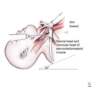
IJ vein landmarks


Image result for right internal jugular vein central line ultrasound

  1. Position patient in trendelenburg position (head angled down toward floor)
    1. Lower risk of air embolism
    2. Engorges vessels and allows for easier visualization
  2. Preparation of the catheter
    1. Flush all three central venous catheter lumens with Normal Saline
    2. Flushing lines is preferred to aspirating as low volume may result in line collapse on aspiration
  3. Preparation of skin
    1. Perform Ultrasound machine preparation and pre-scanning as above
    2. Position head extended and turned away from the insertion site
    3. Apply hibiclens to a wide area over the anterior-lateral neck
    4. Drape the neck to shield all but the prepped skin
  4. Local anesthetic
    1. Clear any air bubbles in a syringe of Lidocaine 1% without Epinephrine
      1. Air bubbles will markedly decrease quality of Ultrasound image
    2. Inject Lidocaine 1% without Epinephrine at the entry site
      1. Raise a skin wheal at insertion site
      2. Infiltrate along expected needle insertion tract
        1. Aspirate prior to injecting to prevent intravascular injection
  5. Needle insertion site
    1. Use Ultrasound localization technique described above
    2. Insertion site
      1. Insertion site will be lateral to palpated carotid pulsation
      2. Approximately at top of triangle formed by sternocleidomastoid muscles bodies and clavicle
      3. Caution
        1. Internal Jugular Vein positioning is variable
        2. Ultrasound guidance is far preferred as landmarks are unreliable
        3. Avoid inserting needle through the sternocleidomastoid muscle (hematoma risk)
      4. Landmark triangle (insertion is at the apex of triangle, where two bodies of SCM meet)
        1. Anterior sternocleidomastoid muscle (SCM) body
        2. Posterior sternocleidomastoid muscle (SCM) body
        3. Clavicle (base of triangle)
      5. Landmarks by finger breadths
        1. Three fingers lateral to midline trachea
        2. Three fingers superior to clavicle (approximate level of cricoid ring)
    3. Needle insertion
      1. Needle types (either is attached to a 10 cc syringe)
        1. Steel Needle 18g (standard, more rigid)
        2. Angiocatheter 18g - long (alternative to steel needle)
          1. Angiocatheter (18 gauge) is typically included in the Central Line kit
          2. Once in lumen, remove needle and thread wire through catheter
          3. May be easier to maintain catheter within vessel lumen while threading guide wire
          4. In large patients, angiocatheter may be too short to access the vessel lumen
      2. Needle is directed toward nipple on side of insertion
      3. Insert needle at 45 degrees to the skin plane (when using Ultrasound guidance)
        1. Landmark insertion (without Ultrasound) is typically at a 30 degree angle to the skin plane
      4. Advance needle as described above under technique of Ultrasound-guided needle insertion
        1. Internal jugular is typically superficial (2-3 cm depth from skin surface)
        2. Aspirate while inserting needle
        3. Advance the needle another 0.5 cm past the time blood is first aspirated (to ensure in lumen)
  6. Guide-wire insertion
    1. Remove syringe from needle
    2. Occlude the open needle base to prevent bleeding and air embolism
    3. Insert guidewire
      1. Some recommend observing guidewire enter vessel on Ultrasound
    4. Typically insert guidewire until free end is approximately at the level of the patient's head
      1. Withdraw guidewire a short distance if ectopy seen on telemetry monitor
  7. Withdraw needle
    1. Firmly grasp guide wire
    2. Back out over the wire
    3. Adjust grasp on wire to be at skin entry site once needle is withdrawn
  8. Make skin nick
    1. Nick skin with #11 blade along the edge of the wire insertion site
    2. Confirm that the nick is contiguous with the space the wire lies within
  9. Dilator insertion
    1. Insert dilator over the wire and into the skin
      1. Do not fully insert dilator
      2. Only insert dilator far enough to dilate skin and soft tissue, but not vessel
    2. Twist the dilator to assist in advancing past resistance
    3. Withdraw the dilator
  10. Central catheter insertion
    1. Always have hold of guidewire throughout this process
    2. Insert catheter over the guide wire via the longest, most distal port (remove brown cap)
      1. As catheter approaches skin, if guidewire does not emerge through port
        1. Withdraw the guidewire from skin until it emerges via port
      2. Grasp the guidewire at the distal port prior to letting go of guidewire at skin
    3. Advance catheter through skin to estimated depth
      1. Err on the side of caution by inserting further than estimate (e.g. 15 cm right, 20 cm left)
        1. Line may be withdrawn if inserted too far
        2. Line may not be inserted deeper after initial placement
          1. Deeper insertion requires replacement of line over another guidewire
      2. Typical final insertion depths (as above, insert further than these depths initially)
        1. Right side: Men 12-13 cm, Women: 11-12 cm
        2. Left side: Add 5 cm to right side length
    4. Remove guidewire
    5. Flush all 3 lines (all three lines should have been filled with saline in preparation)
  11. Confirm catheter placement
    1. Secure Central Line
    2. Portable Chest XRay
      1. Central Line tip should be at superior vena cava junction with right atrium
      2. Approximate tip position is 2 cm below the superior right heart sillhouette
      3. Tip will be 4-5 cm below the carina, just below the hilum
    3. Adjust Central Line based on Chest XRay (may withdraw, but may not insert further due to infection risk)
    4. Suture the Central Line in place

2016年2月17日星期三

study resource for liver disease

http://www.hepatitis.va.gov/provider/guidelines/2009cirrhosis.asp
http://intranet/websitefiles/mmcintranet25168/body.cfm?id=2906 (intranet in montefiore)

2016年2月12日星期五

Food impactions in adult


Introduction: accidental foreign body or large food bolus ingestion in children, or mentally impaired older adults
Food bolus (typically meat) impaction above pre existing esophageal stricture or ring is by far the most common cause of esophageal body obstruction
By comparison, foreign body is the MCC in children (coins)

More than 80% ingested foreign bodies pass without the need of intervention.
In the setting of intentional ingestions, endoscopic intervention is required in up to 76% of patients, and surgical intervention is required up to 16%

Complications: ulcer formation, laceration, perforation, intestinal obstruction, aortoesophageal fistula formation, tracheoesophageal fistula formation and bacteremia
Food impaction often happen in physiologic or pathologic luminal narrowing area:
- upper esophageal sphincter
- level of aortic arch
- diaphragmatic hiatus

Structural or functional esophageal abnormalities that increase the risk of foreign body/food impaction in the esophagus include diverticula, webs, rings, strictures, achalasia, and tumors

Clinical presentation:

Acute onset of dysphagia or complete inability to swallow saliva
92% dysphagia, 60% neck tenderness
Inability to swallow oral secretions is an important symptom which indicates total obstruction

Others: choking, refusal to eat, hypersalivation/drooling, retrosternal fullness, regurgitation of undigested food, wheezing, respiratory distress, odynophagia (indicate laceration or perforation)

Symptoms in patients with a perforation will depend upon the site of the perforation.

Perforation in the oropharynx or proximal esophagus may cause neck swelling, tenderness, erythema, or crepitus.

In the mid or distal esophagus may result in severe retrosternal chest and/or upper abdominal pain, tachypnea, dyspnea, cyanosis, fever, and shock.

Perforation of the stomach, small bowel, or colon may present with signs of peritonitis, such as abdominal pain, rebound, guarding, tachycardia, hypotension, and fever.

Diagnosis:

Radiographic imaging:

Plain neck, chest, and abdominal radiographs may reveal a radiopaque foreign body or signs of esophageal perforation

Computed tomographic (CT) scanning may be helpful if plain radiographs are negative, particularly in patients suspected of having ingested packets of narcotics or other drugs

Examinations using oral contrast, such as a barium swallow, should not be performed, since contrast administration may impair subsequent endoscopic visualization.

Management:

Conservative management is appropriate for the majority of patients, since most objects will pass uneventfully

Timing of endoscopy — Patients requiring endoscopy can be triaged into one of three groups: those requiring emergent endoscopy, those requiring urgent endoscopy(within 24 hrs), and those requiring nonurgent endoscopy
 
 
Foreign bodies that have passed into the stomach — Most foreign bodies that enter the stomach will pass in four to six days, and conservative management is appropriate for most blunt objects in asymptomatic patients. As noted above, exceptions include disk batteries, magnets, objects longer than 6 cm, and objects with a diameter >2.5 cm.
Airway management — Airway protection is important for all patients undergoing endoscopic foreign body removal. Oropharyngeal suction is required to avoid pulmonary aspiration. Patients with impactions in the upper esophagus may require endotracheal intubation to protect the airway. The use of an overtube should also be considered to prevent an object from accidentally being dropped into the patient's airway. In addition, a laryngoscope should be immediately available in the event of airway obstruction.
Equipment:
Choice of endoscope — The forward-viewing flexible endoscope has become the instrument of choice in managing foreign bodies in most medical centers because it permits safe extraction of the object and inspection of the esophageal mucosa. Rigid endoscopy may be required to remove foreign bodies in the upper esophagus.
Both flexible and rigid endoscopic approaches are successful in more than 90 percent of cases, but rigid endoscopy is associated with a higher perforation rate.
Management based upon the type of ingestion
Food bolus: The American Society for Gastrointestinal Endoscopy guidelines suggest that food boluses that are not causing complete obstruction be removed within 24 hours. However, we attempt to remove such boluses within 12 hours in order to avoid pulmonary aspiration.
Administration of glucagon (1 mg IV) can be attempted to relax the esophagus, which may promote passage of the food bolus.
Blunt objects: Blunt objects should be removed with equipment that is suited to the shape of the object. Blunt objects that have already entered the stomach can usually be managed conservatively.
Long objectsLong objects (longer than 6 to 10 cm) are unlikely to pass the duodenal sweep and should be removed.
Sharp-pointed objectsThe presence of sharp-pointed objects (such as chicken and fish bones, straightened paper clips, toothpicks, needles, bread-bag clips, and dental bridgework) in the esophagus represents a medical emergency because of the risk of perforation.
Disk batteriesDisk batteries in the esophagus should be removed promptly. Contact of the flat esophageal wall with both poles of the battery conducts electricity that may rapidly result in liquefaction necrosis and perforation
MagnetsIngested magnets may result in severe gastrointestinal injury because the attractive force between magnets or between a magnet and an ingested metal object can trap a portion of the bowel wall and cause necrosis. This can result in fistula formation, perforation, obstruction, volvulus, or peritonitis.
Drug packetsDrug packets ingested by drug traffickers in an attempt to conceal their possession should not be removed endoscopically because of the risk of rupture.
Foreign bodies in the small bowelDeep small bowel enteroscopy has been used as an alternative to surgery for the management of patients with foreign bodies in the small bowel.


Mauriac syndrome


Mauriac first defined glycogenic hepatopathy (GH) in 1930 in a child with brittle diabetes, as a component of Mauriac syndrome, characterized by delayed development, hepatomegaly, cushingoid appearance, and delayed puberty.


From hepatology image of the month in Oct 23 2015 by Seth Sweetser MD from mayo clinic:

A 27-year-old man with poorly controlled type 1 diabetes mellitus (average hemoglobin A1c of 15%) presented with a 1-week history of progressive pressure-like right upper abdominal discomfort associated with early satiety and nausea. On physical exam, he had firm hepatomegaly extending into the right pelvis. Laboratory testing revealed an aspartate aminotransferase = 6720 U/L (normal, 8–43 U/L), alanine aminotransferase level = 2549 U/L (normal, 7–45 U/L), alkaline phosphatase = 529 U/L (normal, 41–108 U/L), total bilirubin = 1.7 mg/dL (normal 0.1–1.0 mg/dL), with direct bilirubin = 1.5 mg/dL (normal 0.0–0.3 mg/dL) and a normal international normalized ratio. A computed tomography (CT) scan of the abdomen showed massive hepatomegaly of increased density as compared to the spleen (Fig. 1). Infectious and autoimmune causes of liver disease were excluded by laboratory testing.
 
A liver biopsy was obtained and revealed preserved parenchymal architecture and enlarged pale hepatocytes (Fig. 2) with abundant cytoplasmic glycogen deposits demonstrated by periodic acid-Schiff stain (Fig. 3) and diastase digestion removing the glycogen resulting in “ghost cells” (Fig. 4). These histologic findings are characteristic of glycogenic hepatopathy.  


History of poorly controlled DM, acute liver injury (marked elevation in aminotransferases and characteristic histologic changes on liver biopsy are diagnostic of glycogen hepatopathy (GH)
The other main cause of liver enlargement and deranged liver tests related in diabetes mellitus is fatty liver.
Fatty liver
Glycogenic hepatopathy
hyperinsulinemia
Insulin deficiency
Mild elevation in liver enzymes
Marked elevation in liver enzymes
Hypodense liver on CT
Hyperdense on CT, bright liver on CT scan without contrast can be the clue
Possible pathogenesis:
 
Hyperglycemia and overinsulinization are believed to be metabolic preconditions for hepatic glycogen accumulation in GH. Hyperglycemia activates glycogen synthase by inhibiting glycogenesis via glycogen phosphorylation inactivation. Glycogen accumulation further increases because insulin also activates glycogen synthase.
Hepatic glycogen accumulation occurs despite the high cytoplasmic glucose concentration in the presence of insulin. Therefore, frequent hyperglycemic episodes and the following insulin therapies
are believed to be the primary pathogenetic mechanisms of hepatomegaly and liver function disorder that develop in type 1 diabetic patient due to glycogen accumulation.
 
Treatment:
GH therapy is performed via establishing glycemic control. Tight glycemic control, providedvia intensive insulin therapy, results in full remission of clinical, laboratory, and histologic abnormalities


2014年8月3日星期日

ccb overdose

CCB overdose -diltiazem and verapamil are the most cardiotoxic of CCB -5-10 times of usual dose may develop severe intoxication, such as drowsiness and confusion, vs may include hypotension and bradycardia, JVD, pulmonary crackles and other sign of heart failure -ECG changes PR interval prolongation and any bradydysrrhythmia finger stick may reveal hyperglycemia treatment: GI decontamination, including gastric lavage IV calcium, glucagon, high dose insulin therapy, lipid emulsion transvenous pacemaker, intraaortic balloon pump and extracorporeal membrance oxygenation for severe poisoned. 1, assess and stabilize airway, breathing and circulation 2, iv crystalloid, isotonic saline 500 to 1000ml boluses 3, bradycardia treat w/ atropine, repeat for 3 total doses 4, iv calcium for hypotension and/or bradycardia, bolus, calcium chloride 10-20ml of 10% solution via central line, calcium gluconate 30-60 ml of 10% solution cont infusion of 0.5 meq calcium/kg per hour and monitor serum calcium and ECG for evidence of hypercalcemia 5, glucagon for bradycardia, bolus therapy 1-5mg iv push, may repeat up to 15mg 6, vasopressor, norepinephrine 2mcg/min iv, titrate rapidly to systolic blood pressure 100mg 7, bolus insulin 1unit/kg iv, dextrose 25-50g iv, repeat for hypoglycemia, give potassium for hypokalemia 8, GI decontamination: activated charcoal 1g/kg up to 50g 9, consider following therapies if above fail: transvenous cardiac pacing, intraaortic balloon pump, cardiopulmonary bypass, extracorporeal membrane oxygenation

2013年11月18日星期一

IPPF very rare interstitial peumonia

Idiopathic pleuroparenchymal fibroelastosis

Clinical Features:

Patient 1 was a 65-year-old woman with “incidental” pleural thickening seen on a chest radiograph 10 years prior to referral. Four years prior to referral, she underwent a modified radical mastectomy for adenocarcinoma of the breast and was treated with adjuvant chemotherapy of cyclophosphamide, methotrexate, and fluorouracil (CMF) without radiation therapy. The patient was treated with daily doses of tamoxifen, and she returned to her baseline function of aerobic exercise 60 min daily. Eight months prior to referral, dyspnea on exertion and a nonproductive cough developed. Chest radiography revealed a new right pleural effusion and progressive, pleural thickening (greater in the right lung than in the left) when compared to prior radiographs. Pleural fluid was negative for malignancy. A diagnostic video-assisted thorascopic lung biopsy was performed, and at surgery a complex pleural process with extensive fibrosis requiring decortication was found. Physical examination findings were significant for tachypnea, tachycardia, diffusely decreased breath sounds, and decreased diaphragmatic excursion. Extensive evaluation was negative for pneumoconiosis, autoimmune disease, and HLA-B27–related disease.

Patient 2 was a 52-year-old man in whom exertional breathlessness developed following an upper respiratory tract infection. A chest radiograph revealed bilateral pleural thickening. One year later, he underwent right-sided pleural decortication with symptomatic improvement. However, several months later, progressive dyspnea again developed. Ultimately, the patient underwent left pleural decortication. The patient returned to work but remained dyspneic with a productive cough. He was then referred to our institution. His medical history was significant for Charcot-Marie-Tooth disease (hereditary motor sensory neuropathy), gastroesophageal reflux, and narcolepsy. A sister had sarcoidosis, and he had previously worked in both construction and agriculture, although a direct exposure to asbestos was absent. On examination, he was mildly tachycardic and tachypneic. Fine mid-late inspiratory rales and distant breath sounds were noted. Autoimmune serology findings were negative.

Patient 3 was a 61-year-old woman who presented with chronic cough. Diagnosed with asthma in her 20s, she had experienced multiple episodes of “bronchitis” and “pneumonia” over her lifetime. At the time of referral, the cough was accompanied by wheezing, dyspnea, and chest tightness that occurred on a daily basis. Her medical history was significant for breast cancer that had been diagnosed and treated 10 years previously with a left modified radical mastectomy, chemotherapy with CMF, and radiation therapy. She had experienced no significant environmental or occupational exposures. Examination findings noted mild tachypnea and tachycardia with fine mid-late inspiratory rales at the left base, and mild-moderate kyphoscoliosis, and a mid-systolic click found during her cardiologic examination that was consistent with mitral valve prolapse. Bronchoscopy, routine laboratory examination findings, autoimmune serology results, cystic fibrosis genotyping results, and α1-antitrypsin levels were all unrevealing. Prior chest radiographs and chest CT scans had revealed biapical pleural thickening 5 years earlier.

Patient 4 reported reduced exercise tolerance as a teenager. At age 32 years, she underwent a pulmonary evaluation following a diagnosis of pulmonary fibrosis in her twin sister. Her pulmonary physiology was restrictive, but as she was asymptomatic no further evaluation was performed. Seven years later, cough and pleuritic chest pain developed. An examination revealed a loud pleural rub, and she was treated with antibiotics. Over the next year, recurrent pneumonias developed and she was referred to our institution. The patient was a life-long nonsmoker without occupational or environmental exposures. Family history was significant for a twin sister who died of progressive pleuroparenchymal fibrosis (ie, patient 5) and a grandfather with rheumatoid arthritis. An examination revealed mild tachypnea and tachycardia, a pleural rub at the right base, and rales at the left base. Laboratory examination findings were notable for an erythrocyte sedimentation rate of 47 mm/h but negative autoimmune serology results.

Patient 5, the twin sister of patient 4, presented in 1987 at age 32 years with right upper lobe pneumonia. Chest radiographs revealed progressive upper lobe volume loss and bilateral pleural thickening. Her pulmonary physiology was restrictive. The patient was a nonsmoker and worked as a nurse. Her anti-nuclear antibody titer was 1:80, and rheumatoid factor titer was 1:160 without other evidence of a connective tissue disease. An evaluation for HLA-B27–associated disease was negative. The patient underwent surgical lung biopsy that showed pleural fibrosis and patchy interstitial fibrosis. The patient was treated with steroids without improvement. She died of progressive respiratory failure 5 years after undergoing surgical lung biopsy. An autopsy revealed marked pleural and interstitial fibrosis that was identical to that found in her twin sister. In summary, we have reported five cases of a unique idiopathic pleuroparenchymal lung disease that is characterized by upper lobe radiographic predominance and pathologic findings that do not fit with any of the currently defined interstitial pneumonias. We term this disorder idiopathic pleuroparenchymal fibroelastosis.

cite from http://www.ncbi.nlm.nih.gov/pubmed/23169023

Case 1 chest CT: biapical fibrotic changes, pleural and parenchymal densities in the right upper lobe as well as mild emphysematic changes.

Case 2 pathology: hematoxylin-eosin stain showing abundance of elastic fibers in addition to collagen fibers and abrupt transition from fibroelastosis to unaffected normal lung parenchyma. Insert: slide 'thumbnail' view demonstrating an extraordinarily thick pleural cap with sparing of the adjacent lung parenchyma.


2013年10月30日星期三

gastroenterology ob 6

Esophagitis dissecans superficialis


 2009 Dec;33(12):1789-94. doi: 10.1097/PAS.0b013e3181b7ce21.

Esophagitis dissecans superficialis ("sloughing esophagitis"): a clinicopathologic study of 12 cases.

Source

Department of Pathology, VA North Texas Health Care System, The University of Texas Southwestern Medical Center, Dallas, USA.

Abstract

Esophagitis dissecans superficialis (EDS) is a term applied to a rare endoscopic finding characterized by sloughing of large fragments of the esophageal squamous mucosa that may be coughed up or vomited. Although EDS has been reported in association with certain medications and esophageal strictures, most cases remain unexplained and the histopathologic features of EDS are inadequately described. We undertook this study to define useful diagnostic criteria based on the examination of a series of well-characterized cases of EDS. To identify patients with EDS, we searched our endoscopy and pathology databases, reviewed the esophageal biopsy specimens from candidate cases, and correlated them with pertinent clinical information. Twelve patients (11 men and 1 woman) had endoscopic and histologic findings of EDS and 9 had the histologic features without the endoscopic correlates. Biopsies from confirmed EDS patients showed sloughing and flaking of superficial squamous epithelium with occasional bullous separation of the layers, parakeratosis, and varying degrees of acute or chronic inflammation. Fungal elements were identified in 3 patients, but were not associated with acute inflammation. None of the EDS patients were on bisphosphonate therapy or had bullous skin disorders. Follow-up endoscopy in 5 patients showed complete resolution of the esophageal abnormalities in 4 and mild esophagitis in one. In spite of its sometimes, dramatic presentation, EDS is a benign condition that resolves without lasting esophageal pathology. Although an association with medications, skin conditions, heavy smoking, and physical trauma has been reported, the pathogenesis of EDS remains unexplained.

Endoscopic view of esophagitis dissecans superficialis. A: With diffuse sloughing mucosa of the lower esophagus in a 76-year-old woman presenting hematemesis, and the cause was idiopathic; B: with longitudinal sloughing mucosa from upper to mid esophagus in a 67-year-old woman with mucocutaneous type pemphigus vulgaris, note fine whitish fragments of sloughed mucosa, and the index value for anti-desmoglein 3 antibody by enzyme-linked immunosorbent assay was over 1280 (normal value < 7).


PANCREATIC REST — A pancreatic rest (also known as ectopic pancreas, aberrant pancreas, and heterotopic pancreas) refers to ectopic pancreatic tissue. These rare submucosal tumors most commonly consist of cystically dilated exocrine cells. Endocrine pancreatic tissue or a combination of exocrine and endocrine cell types may also be seen.
Pancreatic rests are most frequently found in the distal stomach, duodenum, or proximal jejunum, but have also been reported within a Meckel's diverticulum, the gallbladder, bile ducts, and the minor and major papillae. They are typically discovered incidentally during endoscopy, surgery, or autopsy. They are also occasionally found on CT scan. CT findings that may help differentiate pancreatic rests from other submucosal lesions identified in one study included:
  • A flat-ovoid shape (long diameter to short diameter ratio of greater than 1.4)
  • Location of the lesion in the antrum, pylorus, or duodenum
  • An endoluminal growth pattern
  • An ill-defined border
  • Prominent enhancement of the overlying mucosa
The study found that the presence of at least two of the above findings had a sensitivity of 100 percent and a specificity of 82.5 percent for diagnosing a pancreatic rest in the upper gastrointestinal tract [52]. The specificity increased to 100 percent if three of the above findings were present.
However, while pancreatic rests may be detected with CT scan, if a submucosal lesion is noted on upper endoscopy, we suggest endoscopic ultrasound with endoscopic mucosal resection for further evaluation, as small lesions may be missed on CT scan. (See 'General principals of tissue sampling' above.)
Complications of pancreatic rests are rare, but may include ulceration, gastric outlet obstruction, and malignancy [50,53].
Endoscopic appearance — A pancreatic rest appears as a submucosal nodule, usually with a central umbilication that corresponds to a draining duct.
Endosonographic findings — Pancreatic rests are hypoechoic or intermediate echogenic heterogeneous lesions with indistinct margins. They most commonly arise from the third or fourth layer, or a combination of the two layers of the GI tract. Anechoic areas within the lesion correlate with ductal structures.
Diagnosis and treatment — The diagnosis can be made histologically from tissue obtained by biopsy forceps or snare excision, although techniques to obtain deeper biopsies (using jumbo biopsy forceps, tunnel biopsy, endoscopic mucosal resection, or EUS-guided FNA) may be required. The management strategy should be guided by symptoms and suspicion for malignancy. Asymptomatic lesions can be followed expectantly. Endoscopic resection can be performed by standard snare, band ligation-assisted, or cap-assisted polypectomy technique. Surgical resection is preferred to endoscopic resection when the muscularis propria is involved.

Endoscopic photograph of a "dimpled" antral mass in an 8 yo found incidentally.
Endoscopic photograph of a "dimpled" antral mass in a 5 yo found incidentally


                                                                          Fundoplication